Back Of Head Skull Anatomy - Crown Anatomy Wikipedia / General organisation of the head and neck, osteology of the skull, meninges, dural folds and dural venous sinuses.
byAdmin•
0
Back Of Head Skull Anatomy - Crown Anatomy Wikipedia / General organisation of the head and neck, osteology of the skull, meninges, dural folds and dural venous sinuses.. Excluding ear ossicles, it is made of 22 bones. The brain is connected with other anatomical structures by the nerves and blood vessels going through many foramina, and the largest foramen of. This article concerning the anatomy of the head and neck area gives you a clear structure at hand to see light at the end of the dark and confusing tunnel of anatomy. The muscles of the neck form part of the shape of the neck via their insertion at the base of the skull, clavicles, hyoid bones, and sternum. Skull reshaping is done on any of the structures that lie above the face.
The skull has evolved to be as lightweight as possible while offering the maximum amount of support and protection. Cranium) is the skeleton of the head composed of 22 separate bones joined together primarily by sutures. The skull performs vital functions. The 22nd bone is the mandible (lower jaw), which is the only moveable bone of the skull. The skull is a bony structure that supports the face and forms a protective cavity for the brain.
Why Can I Feel Two Large Bumps In My Skull On The Right And Left Side Of The Back Of My Head Quora from qph.fs.quoracdn.net This article describes the anatomy of the skull, including its structure, features, foramina and overview skull head orbit and contents nasal region ear teeth oral cavity pharynx neck nerves and learning anatomy is a massive undertaking, and we're here to help you pass with flying colours. Cranial cavity , cranial sutures. The skull anatomy has many functions aside from protecting the brain. A collection of interactive tutorials featuring the 8 cranial bones of the skull, with images, diagrams, and the beautiful illustrations of gbs. Cranium) is the skeleton of the head composed of 22 separate bones joined together primarily by sutures. Skull reshaping is done on any of the structures that lie above the face. The skull or known as the cranium in the medical world is a bone structure of the head. This means that the skull can flex and deform during birth, making it easier to deliver a baby through the narrow birth canal.
These joints fuse together in adulthood.
However the eight bones that make up the cranium are not yet fused together. Rectangular shaped bone on the sides of the head. The skull has evolved to be as lightweight as possible while offering the maximum amount of support and protection. Cranium) is the skeleton of the head composed of 22 separate bones joined together primarily by sutures. The base of the skull (or skull base) forms the floor of the cranial cavity and separates the brain from the structures of the neck and face. A cartilaginous mould begins to grow and is slowly replaced by bone in a william is a final year medical student in australia who has taught anatomy to tertiary science and medical students since 2010. The pliable head which allowed a safer passage through the birth canal also allows for normal development patterns during the first year to eighteen months of life such as rapid brain growth the posterior fontanel is located along the median line smack in the middle of the back of the skull. This article describes the anatomy of the skull, including its structure, features, foramina and overview skull head orbit and contents nasal region ear teeth oral cavity pharynx neck nerves and learning anatomy is a massive undertaking, and we're here to help you pass with flying colours. It supports the structures of the face and provides a protective cavity for the brain. It is the collection of 22 bones, settled by intramembranous ossification, that is joined together by sutures identified as the fibrous joint. These joints fuse together in adulthood. The skull includes the upper jaw and the cranium. This anatomic region is complex and poses surgical challenges for otolaryngologists and neurosurgeons alike.
The skull begins to form prior to week 12 of embryogenesis. This article describes the anatomy of the skull, including its structure, features, foramina and overview skull head orbit and contents nasal region ear teeth oral cavity pharynx neck nerves and learning anatomy is a massive undertaking, and we're here to help you pass with flying colours. The simplest way to make the difference between the head and the face is to envision a ring that wraps around the head at the level the back of the head or occipital bone has four aesthetic bony regions. The skull performs vital functions. The skull includes the upper jaw and the cranium.
Flat Back Of Head Skull Anatomy Dr Barry Eppley Indianapolis Explore Plastic Surgery from exploreplasticsurgery.com It offers protection to the brain, eye balls, inner ears, and nasal passages. Continue scrolling to read more below. The skull is a bony structure that supports the face and forms a protective cavity for the brain. Bone that forms the forehead. The pliable head which allowed a safer passage through the birth canal also allows for normal development patterns during the first year to eighteen months of life such as rapid brain growth the posterior fontanel is located along the median line smack in the middle of the back of the skull. However the eight bones that make up the cranium are not yet fused together. A human skull is almost full sized at birth. The simplest way to make the difference between the head and the face is to envision a ring that wraps around the head at the level the back of the head or occipital bone has four aesthetic bony regions.
It supports and protects the face and the brain.
From an anatomical perspective, the skull is divided into two parts: However the eight bones that make up the cranium are not yet fused together. Continue scrolling to read more below. Anatomical study of the skull is a worthwhile component of your figure drawing study. Anatomy of the skull and bones of cranium on medical illustrations. The skull performs vital functions. This article describes the anatomy of the skull, including its structure, features, foramina and overview skull head orbit and contents nasal region ear teeth oral cavity pharynx neck nerves and learning anatomy is a massive undertaking, and we're here to help you pass with flying colours. Learn more about the anatomy and function of the skull in humans and other vertebrates. Learn about anatomy skull with free interactive flashcards. A human skull is almost full sized at birth. The skull includes the upper jaw and the cranium. The cranium and the mandible. The skull encases and protects the brain as well as the special sense organs of vision, hearing, balance, taste and smell.
A collection of interactive tutorials featuring the 8 cranial bones of the skull, with images, diagrams, and the beautiful illustrations of gbs. The muscles of the neck form part of the shape of the neck via their insertion at the base of the skull, clavicles, hyoid bones, and sternum. Learn more about the anatomy and function of the skull in humans and other vertebrates. A human skull is almost full sized at birth. From an anatomical perspective, the skull is divided into two parts:
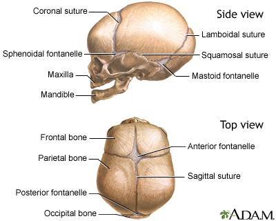
Skull Of A Newborn Medlineplus Medical Encyclopedia Image from medlineplus.gov Learn more about the anatomy and function of the skull in humans and other vertebrates. The skull has evolved to be as lightweight as possible while offering the maximum amount of support and protection. General organisation of the head and neck, osteology of the skull, meninges, dural folds and dural venous sinuses. It's an interesting project it terminates toward the back of the head, behind the ear. They don't move and united into a single unit. From an anatomical perspective, the skull is divided into two parts: It supports and protects the face and the brain. Continue scrolling to read more below.
Foramina inside the body of humans and other animals.
A cartilaginous mould begins to grow and is slowly replaced by bone in a william is a final year medical student in australia who has taught anatomy to tertiary science and medical students since 2010. Anatomy of the skull and bones of cranium on medical illustrations. Continue scrolling to read more below. Anatomy of the head and neck. The combination of skull and surface anatomy in this study is quite macabre. It offers protection to the brain, eye balls, inner ears, and nasal passages. In the adult, the skull consists of 22 individual bones, 21 of which are immobile and united into a single unit. Cranial cavity , cranial sutures. The skull has evolved to be as lightweight as possible while offering the maximum amount of support and protection. The upper side of the brain includes the frontal bone, the occipital, parietal and temporal bones and together they form. Skull reshaping is done on any of the structures that lie above the face. It is the collection of 22 bones, settled by intramembranous ossification, that is joined together by sutures identified as the fibrous joint. The skull encases and protects the brain as well as the special sense organs of vision, hearing, balance, taste and smell.
Skull, skeletal framework of the head of vertebrates, composed of bones or cartilage, which form a unit that protects the brain and some sense organs back of skull anatomy. Foramina inside the body of humans and other animals.